This content is for informational purposes only and does not constitute medical advice. Please consult with a licensed podiatrist for a personalized evaluation and treatment plan. Individual results may vary.

A foot or ankle fracture involves a partial or complete break in the bone, resulting from high-impact trauma, rotational injuries, or repetitive mechanical stress. Primary symptoms include immediate, localized pain, significant ankle injury swelling, visible bruising, and a functional inability to support weight on the affected limb. Immediate clinical evaluation is required if the injury presents with a physical deformity, skin penetration by bone fragments, or neurological symptoms such as numbness or tingling. Early intervention by a specialist is essential to prevent malunion, chronic joint instability, or the rapid onset of post-traumatic osteoarthritis.

For patients in Southern California seeking expert diagnosis and a personalized recovery roadmap, consulting an experienced podiatrist in LA like Dr. Arkady Kaplansky provides access to state-of-the-art surgical and regenerative therapies.

Identifying the Signs of a Broken Ankle and Foot

Distinguishing between a severe ligamentous sprain and a bone fracture is critical for establishing an effective fractured foot treatment plan. While pain is a universal indicator of injury, specific clinical markers suggest structural bone failure.

Acute Physical Indicators

The most common signs of a broken ankle or foot include:

  • Audible Trauma: A distinct “cracking” or “snapping” sound at the moment of impact.
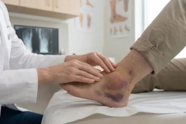
  • Point Tenderness: Intense pain isolated to a specific bony area, such as the lateral or medial malleolus (the protrusions on either side of the ankle).
  • Rapid Edema and Discoloration: Significant ankle injury swelling that occurs within minutes, often followed by deep purple bruising as blood accumulates around the fracture site.
  • Mechanical Instability: A sensation that the joint cannot support the body’s weight, or visible misalignment of the foot relative to the leg.

A softly lit, natural daylight photo captured from a medium side angle. A female podiatrist in a crisp white lab coat sits and uses both hands to perform a careful examination of a bare male foot and ankle, which has significant dark purplish bruising and swelling visible. The patient is seated and looking down. The clean background includes wood-grain cabinetry, a large window providing daylight.
AI-generated for demonstration

Stress Fractures vs. Traumatic Breaks

Unlike acute fractures caused by a single event, stress fractures are microscopic cracks resulting from chronic overuse. These are particularly common among Los Angeles athletes and long-distance runners. While the symptoms may start as a dull ache, they progress to sharp pain that persists even during rest, eventually requiring a specialized foot fracture boot to allow the bone to remodel.

Diagnostic Standards: The Role of Imaging

Accurate ankle break treatment begins with precise visualization of the injury. A clinical examination assesses the neurovascular status of the foot, ensuring that blood flow and nerve function remain intact. However, definitive diagnosis relies on high-resolution imaging.

  1. X-ray of a Broken Ankle: This is the foundational diagnostic tool used to identify the location, direction, and type of fracture (e.g., transverse, oblique, or comminuted).
  2. Computed Tomography (CT) Scans: Often utilized for intra-articular fractures (breaks that extend into the joint), a CT scan provides a three-dimensional view of the bone fragments, which is vital for an orthopaedic surgeon planning a complex reconstruction.
  3. Magnetic Resonance Imaging (MRI): While X-rays show the bone, an MRI reveals associated damage to the syndesmosis ligaments, tendons, and cartilage that often accompanies high-energy ankle breaks.

Advanced Surgical and Regenerative Treatment Options

Modern podiatric medicine has moved beyond simple casting. To ensure a faster and more complete broken ankle recovery, a combination of surgical precision and biological acceleration is employed.

Foot and Ankle Surgery (ORIF)

For fractures that are displaced or unstable, Foot and Ankle Surgery is the gold standard. The procedure, known as Open Reduction Internal Fixation (ORIF), involves the surgical realignment of the bone fragments. The surgeon utilizes medical-grade titanium plates, screws, or intramedullary nails to secure the bones in their correct anatomical position. This rigid internal fixation allows for earlier mobilization, which is key to preventing joint stiffness and muscle atrophy.

Platinum Biologics for Bone Healing

To augment the body’s natural healing capacity, Platinum Biologics are integrated into the treatment plan. This advanced therapy includes the use of Platelet-Rich Plasma (PRP) or concentrated bone marrow aspirate. By injecting these high-potency growth factors directly into the fracture site, the recruitment of osteoblasts (bone-building cells) is significantly accelerated. This is particularly beneficial for patients with compromised healing factors or those with high-demand lifestyles who require a swift return to activity.

Remy Laser Pain Treatment

Pain management is a critical component of the recovery phase. Remy Laser Pain Treatment utilizes high-intensity laser therapy to address both acute post-traumatic pain and post-surgical inflammation. The laser energy penetrates deep into the soft tissues to stimulate mitochondrial activity and cellular regeneration. This non-invasive modality reduces the need for systemic pain medications and helps dissipate the residual ankle injury swelling that often lingers after a major break.

Post-Traumatic Rehabilitation and Long-Term Support

The success of fractured foot treatment is not determined solely by the initial healing of the bone, but by the restoration of the patient’s natural gait and balance.

Specialized Wound Care

For patients with open fractures or those recovering from surgical intervention, meticulous Wound Care is paramount. Preventing infection and ensuring that surgical incisions heal without excessive fibrotic tissue (scarring) is essential for maintaining the elasticity of the skin and underlying fascia. Clinical wound management ensures that the barrier remains intact and the recovery is not sidelined by secondary complications.
A natural-light photograph from a low side angle, looking slightly up at a male patient seated on a clean leather examination table. He wears a heavy, black rigid medical walking boot (fracture boot) with straps. The patient looks down at his boot with a contemplative expression. The background features wood-grain cabinetry and a large digital wall monitor clearly displaying a high-contrast X-ray of a human foot and ankle (the word "X-RAY" is visible on screen but blurred). A healthcare provider’s lower body is partially in the foreground. Bright, clear clinic interior.
AI-generated for demonstration

Biomechanical Stabilization with Foot Orthotics

Once the fracture has achieved clinical union, the transition back to standard footwear requires careful management. Custom Foot Orthotics are frequently prescribed to provide structural support during the final stages of broken ankle recovery. These medical devices are engineered to:

  • Stabilize the subtalar and talocrural joints.
  • Redistribute pressure away from the former fracture site.
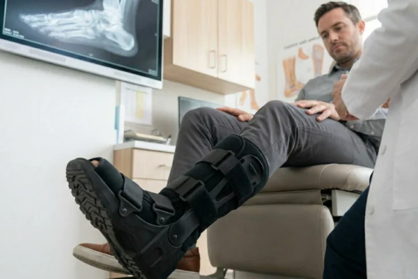
  • Correct compensatory gait patterns that may have developed during the weeks spent in a foot fracture boot.
Treatment Phase Primary Objective Key Modalities
Acute Phase Stabilization & Pain Control Casting, Splinting, Remy Laser
Surgical Phase Anatomical Realignment ORIF Surgery, Platinum Biologics
Sub-Acute Phase Bone Consolidation NWB (Non-Weight Bearing), Wound Care
Remodeling Phase Functional Restoration Physical Therapy, Custom Orthotics

Navigating the Recovery Timeline in Los Angeles

The duration of broken ankle recovery varies based on the severity of the break and the patient’s overall health. Generally, primary bone union occurs within six to eight weeks. However, the remodeling phase—where the bone regains its full strength—can take up to a year.

In a metropolis like Los Angeles, where physical activity and mobility are central to daily life, a multidisciplinary approach is vital. Utilizing an orthopaedic surgeon who integrates regenerative medicine with traditional surgical techniques ensures that the bone does not just “close,” but returns to its peak functional state.

Neglecting early broken ankle symptoms can lead to permanent joint degeneration. Seeking professional intervention at the onset of injury is the most effective way to ensure long-term mobility and prevent the chronic pain associated with improperly healed fractures.

Frequently Asked Questions

How can I tell the difference between a severe sprain and a broken ankle?

While both injuries cause pain, broken ankle symptoms usually include “point tenderness” directly over the bone and a total inability to bear weight. If you notice rapid ankle injury swelling or a visible deformity, a professional clinical evaluation and an x-ray of a broken ankle are necessary to confirm the diagnosis.

Is it possible to walk on a fractured foot or ankle?

While some minor stress fractures allow for limited movement, walking on a displaced break can cause further structural damage and significantly delay broken ankle recovery. Most patients require immobilization in a foot fracture boot or cast to ensure the bones remain in anatomical alignment during the critical healing phase.

Does every foot or ankle fracture require surgical intervention?

Not all injuries require foot and ankle surgery; many stable fractures are successfully managed with non-surgical stabilization and rest. However, if the bone is displaced or involves a joint surface, an orthopaedic surgeon may perform an ORIF procedure to restore alignment and prevent long-term joint degeneration.

What is the typical timeline for fractured foot treatment and recovery?

Most bones require six to eight weeks to achieve primary union, though the full remodeling process can take several months. Advanced modalities like Platinum Biologics and Remy Laser Pain Treatment are often used to accelerate cellular regeneration and manage post-traumatic inflammation during this period.

Why are custom foot orthotics recommended after a fracture heals?

After completing fractured foot treatment, the foot’s internal mechanics may change, leading to new pressure points or gait imbalances. Custom foot orthotics provide essential long-term support, redistributing weight and protecting weakened joints from secondary injuries as you transition back to regular footwear.