This content is for informational purposes only and does not constitute medical advice. Please consult with a licensed podiatrist for a personalized evaluation and treatment plan. Individual results may vary.
Cavus foot is a structural condition where the foot arch is abnormally high. The arch remains elevated even during weight-bearing activities. This layout places immense pressure on the heel and the ball of the foot. Over time, individuals experience chronic arch pain in feet. This imbalance often leads to metatarsalgia, calluses, and severe ankle instability. Early medical evaluation prevents permanent structural damage. Persistent aching arches or recurrent ankle sprains require a professional assessment.
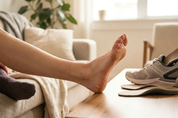
Proper lower limb alignment is essential for balanced mobility. Residents of Southern California can find expert care at a Los Angeles podiatry center. A specialist Dr. Arkady Kaplansky can design a personalized treatment plan for structural relief.
Understanding the Biomechanics of High Arches
A standard foot arch acts as a natural shock absorber. The arch flattens slightly during impact to distribute body weight evenly. A high arch foot lacks this vital flexibility. The middle section of the foot does not touch the ground properly. Every step concentrates impact forces onto the heel bone and the metatarsal heads.
This poor weight distribution alters the standard gait cycle. The lower leg often rotates outward to compensate for the rigid arch. This movement pattern is known as supination or underpronation. The mechanical strain travels up the kinetic chain over time. This shift places undue stress on the shins, knees, hips, and lower back.
The rigid framework forces the lower leg muscles to work harder. The tibialis anterior muscle experiences constant strain. This overwork leads to chronic muscular fatigue and localized soft-tissue inflammation.
Primary Causes of a High Arch Foot
Genetic Factors and Inherited Traits
Underlying Neurological Conditions
According to The American College of Foot and Ankle Surgeons , several conditions alter foot architecture. Common neurological causes include:
- Charcot-Marie-Tooth disease weakens peripheral nerves.
- Spina bifida causes localized spinal nerve damage.
- Cerebral palsy affects muscle tone and coordination.
- Polio damages motor neurons in the spinal cord.
- Stroke can interrupt motor pathways and cause muscle contractions.
The Pain Spectrum and Common Complications
Plantar Fasciitis
Metatarsalgia and Sesamoiditis
Progressive Toe Deformities
Lateral Ankle Instability
Professional Diagnostic Methodologies
Visual Gait Analysis
Radiographic Imaging Matrix
- Meary’s angle measures the alignment of the talus and first metatarsal.
- The calcaneal pitch angle evaluates the tilt of the heel bone.
Comparative Matrix of Foot Architecture Profiles
| Anatomical Features | High Arch Foot | Neutral Foot Arch | Flat Foot |
|---|---|---|---|
| Arch Height | Exceptionally elevated. | Moderately curved. | Completely collapsed. |
| Shock Absorption | Rigid and inflexible. | Dynamic and flexible. | Overly flexible. |
| Weight Distribution | Heel and ball of foot. | Even across the sole. | Shifts to the inner edge. |
| Gait Pattern | Excessive supination. | Balanced motion. | Excessive overpronation. |
Non-Surgical Treatment Modalities for Mechanical Relief
Advanced Custom Orthotic Engineering
These prescription devices feature specific structural modifications:
- Deep Heel Cups: Designed to stabilize the calcaneus and prevent the heel from sliding or rolling outward.
- Full-Contact Arch Fill: Carefully fills the void beneath the high arch, increasing the foot’s contact surface area to immediately relieve pressure on the heel and forefoot.
- Metatarsal Bars or Pads: Slightly elevates the area just behind the ball of the foot, offloading pressure from painful metatarsal heads.
- Lateral Heel Wedges: A subtle external tilt built into the casing to counteract supination and keep the ankle safely centered.
Specialized Footwear Protocols
- Maximum Midsole Cushioning: Shoes must incorporate advanced shock-absorbing foam materials to compensate for the foot’s lack of natural flexibility.
- Broad, Stable Outsoles: A wider base helps resist the foot’s tendency to roll outward, lowering the risk of ankle sprains.
- High-Volume Toe Box: Extra vertical and horizontal space prevents friction along the tops of claw toes or hammertoes.
- Straight-Lasted Construction: The internal shape of the shoe should be straight rather than curved to properly accommodate a rigid foot structure.
Footwear Feature Matrix
| Recommended Features | Mechanical Purpose | Features to Avoid | Pathological Risks |
|---|---|---|---|
| Thick Midsoles | Absorbs ground shock. | Thin Soles | Increases stress fracture risks. |
| Removable Footbeds | Accommodates custom orthotics. | Narrow Shoe Waists | Causes localized nerve pain. |
| Deep Toe Boxes | Prevents friction sores. | Tapered Toes | Accelerates hammertoe growth. |
Advanced Physical Therapy and Muscle Balancing
Targeted Soft-Tissue Stretching
- Gastrocnemius-Soleus Complex Stretching: Chronic supination keeps the calf muscles tightly contracted. Regular wall stretches or strap-assisted stretches help restore vital ankle flexibility.
- Plantar Fascia Stretching: Crossing the foot over the opposite knee and gently pulling the toes backward helps maintain flexibility in the plantar fascia, lowering the risk of heel spurs.
Lower Extremity Strengthening Regimens
- Peroneal Muscle Strengthening: The peroneal muscles on the outside of the shin are responsible for stabilizing the ankle joint. Utilizing resistance bands to pull the foot outward strengthens these tissues, providing better protection against chronic ankle sprains.
- Proprioceptive Balance Training: Exercising on unstable surfaces, such as foam balance pads or wobble boards, retrains the brain’s positional awareness of the ankle joint, improving overall stability.
Surgical Reconstruction: Realignment Strategies for Advanced Cases
Podiatric surgeons develop personalized surgical plans that combine soft-tissue and bone procedures based on the patient’s specific anatomy:
- Tendon Transfers: Moving a working tendon from one part of the foot to another to rebalance muscle pull, which is particularly effective in neurological cases like Charcot-Marie-Tooth disease.
- Calcaneal Osteotomy: Precisely cutting the heel bone and shifting it inward or outward to correct alignment and stop the ankle from rolling.
- Dorsiflexion Osteotomy of the First Metatarsal: Cutting and elevating a dropped first metatarsal bone to level the forefoot and improve weight distribution.
- Arthrodesis (Joint Fusion): In cases of advanced arthritis or rigid structural deformities, fusing the joints of the midfoot or rearfoot stabilizes the architecture, permanently relieving chronic pain.
Long-Term Maintenance and Structural Prevention
Regular professional podiatric maintenance is essential for safely debriding deep calluses and managing corns without damaging underlying tissues. Additionally, orthotic devices should undergo a clinical evaluation every twelve to eighteen months to ensure the structural shell continues to provide optimal biomechanical support. Proactively managing a high arch helps individuals maintain a healthy, active lifestyle while fully protecting the lower limbs from long-term, debilitating injuries.
Frequently Asked Questions
What are the main symptoms of high arch feet?
High arch feet cause chronic arch pain in feet, painful calluses, and severe metatarsalgia. Individuals also frequently suffer from aching arches, claw toes, and recurrent ankle sprains. These symptoms happen because the rigid foot arch cannot absorb daily ground shock.
Can a high arch foot cause knee or lower back pain?
Yes, an elevated foot arch alters the entire gait cycle. This rigid structure forces the lower leg to rotate outward during motion. Over time, this mechanical strain travels up the kinetic chain to cause knee, hip, and back exhaustion.
What causes pes cavus to develop suddenly?
Sudden development of pes cavus in one foot usually points to an underlying neurological condition. Diseases like Charcot-Marie-Tooth or cerebral palsy cause nerve signals to malfunction. This creates severe muscle imbalances that pull the bones upward.
How do custom orthotics help manage aching arches?
Custom orthotics fill the empty void beneath the high arch structure. They expand the contact surface area to distribute body weight away from pressure points. These prescription devices also feature deep heel cups to stabilize the ankle joint.
Is surgery always necessary for a foot arch injury caused by pes cavus?
No, surgery is typically reserved for severe cases that fail to respond to conservative treatments. Most people find effective relief through custom orthotics, specialized footwear, and targeted physical therapy. If a neurological disease causes progressive deformity, a podiatric surgeon can realign the bones.