This content is for informational purposes only and does not constitute medical advice. Please consult with a licensed podiatrist for a personalized evaluation and treatment plan. Individual results may vary.
Posterior tibial tendon dysfunction (PTTD) is a progressive orthopedic condition characterized by the inflammation, overstretching, or eventual rupture of the primary tendon supporting the medial arch. As the leading cause of an acquired collapsed foot arch in adults, PTTD results in significant inner foot arch pain and a gradual loss of foot stability. Clinical intervention is required when individuals experience persistent swelling along the inner ankle, a visible flattening of the foot, or difficulty performing basic weight-bearing tasks. Early diagnosis is essential to halt the progression toward permanent deformity and secondary ankle arthritis.
For residents in the Los Angeles area seeking specialized care for foot instability or chronic arch discomfort, Dr. Arkady Kaplansky provides comprehensive diagnostic imaging and advanced clinical protocols to stabilize the tendon and restore proper biomechanical function.
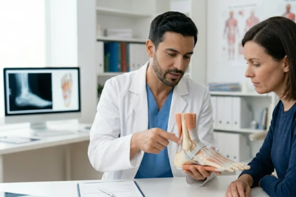
The Biomechanics of Stability: Why the Posterior Tibial Tendon Fails
When this tendon fails, the arch loses its tension, leading to tendonitis foot symptoms that can radiate into the calf and heel. Without this support, the other ligaments are forced to compensate, eventually leading to a structural collapse known as adult-acquired flatfoot. Because this process is often gradual, many patients overlook the early warning signs of posterior tibialis tendinosis until the deformity becomes rigid.
Recognizing the Warning Signs: Symptoms of Posterior Tibial Tendonitis
Clinical Markers of PTTD
- Medial Tenderness: Acute pain along the path of the tendon behind the inner ankle bone.
- The “Too Many Toes” Sign: When viewed from behind, the forefoot of the affected foot rotates outward, making more toes visible on the lateral side than on a healthy foot.
- Localized Edema: Swelling that increases after activity, particularly after standing on the hard surfaces common in urban Los Angeles.
- Single-Limb Heel Raise Failure: An inability to stand on the tiptoes of the affected foot due to the tendon’s inability to stabilize the midfoot.
- Lateral Impingement Pain: As the arch collapses, the heel bone shifts, eventually causing pain on the outside of the ankle where bones begin to rub together.
Clinical Classification: PTTD Stages and Targeted Treatments
PTTD Progression and Service Integration
| Stage | Structural Condition | Foot Deformity | Targeted Clinical Services |
|---|---|---|---|
| Stage 1 | Inflamed; length is normal. | None; arch is intact. | Remy Laser Pain Treatment, NSAIDs. |
| Stage 2 | Stretched or partially torn. | Collapsed foot arch (flexible). | Foot Orthotics, Platinum Biologics. |
| Stage 3 | Severely damaged or non-functional. | Rigid flatfoot; fixed deformity. | Foot and Ankle Surgery, AFO Bracing. |
| Stage 4 | Complete failure; ankle involved. | Advanced arthritis; rigid. | Foot and Ankle Surgery (Complex). |
Advanced Clinical Interventions for Arch Restoration
Remy Laser Pain Treatment: Rapid Inflammation Control
Platinum Biologics: The Regenerative Edge
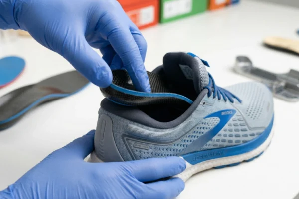
Foot Orthotics: The Gold Standard for Mechanical Support
- Realign the heel bone to reduce the “pull” on the posterior tibial tendon.
- Provide direct mechanical lift to the navicular bone.
- Stabilize the midfoot joints to prevent further arch collapse.
Foot and Ankle Surgery: Structural Reconstruction
Investigating the Root: Posterior Tibial Tendonitis Causes
Predisposing Factors
- Biomechanical Heritage: Individuals born with naturally low arches or “flat feet” place a lifelong, disproportionate strain on the posterior tibial tendon.
- Age-Related Vascularity: As individuals age, the blood supply to the tendon naturally decreases, making the tissue more prone to micro-tears that the body cannot easily repair.
- Obesity and Lifestyle: Increased body mass places exponential force on the arch. Furthermore, occupations in Los Angeles that require long hours of standing on concrete floors are a major contributor to early tendon failure.
- Systemic Health: Conditions like hypertension or diabetes can further compromise the micro-circulation needed for tendon maintenance.
Navigating the Timeline: Posterior Tibial Tendonitis Recovery Time
The Recovery Phases
- Phase 1 (Weeks 1–6): Focus on immobilization and inflammation control. This often involves the use of a walking boot and Remy Laser Pain Treatment sessions to settle the “flare-up.”
- Phase 2 (Weeks 6–12): Transition into weight-bearing. This is when Foot Orthotics are introduced to provide the necessary support as the patient begins walking in traditional footwear again.
- Phase 3 (3–6 Months): Rehabilitation and strengthening. Platinum Biologics may be used during this window to bolster the structural integrity of the healing tissue.
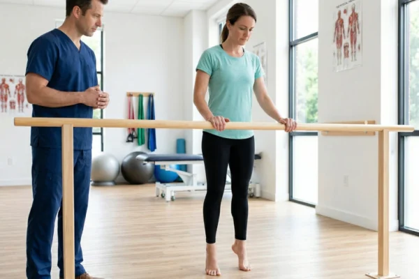
- Phase 4 (6–12 Months): Full return to activity. For those who underwent Foot and Ankle Surgery, this period involves intensive physical therapy to restore the gait cycle and balance.
Differential Diagnosis: Is It PTTD or Something Else?
Comparative Symptom Analysis
| Condition | Primary Pain Location | Distinguishing Feature |
|---|---|---|
| PTTD | Inner ankle and arch. | Arch collapse; inability to do a heel raise. |
| Plantar Fasciitis | Bottom of the heel. | Pain is most severe during the first steps in the morning. |
| Tarsal Tunnel Syndrome | Inner ankle into the toes. | Burning, tingling, or “electric” sensations (nerve-based). |
| Medial Stress Fracture | Directly on the ankle bone. | Constant pain that persists even when not weight-bearing. |
Posterior tibial tendon dysfunction is a progressive condition that, if left unaddressed, can fundamentally alter an individual’s mobility and quality of life. However, with modern interventions such as Foot Orthotics, Remy Laser Pain Treatment, and Platinum Biologics, the transition from a flexible deformity to a rigid, arthritic state is no longer inevitable. By identifying the symptoms of posterior tibial tendonitis early and utilizing a combination of mechanical support and regenerative technology, patients can effectively manage their condition and avoid the complexities of advanced Foot and Ankle Surgery.
Frequently Asked Questions
What are the early symptoms of posterior tibial tendonitis?
Can a collapsed foot arch be corrected without surgery?
What are the primary posterior tibial tendonitis causes?
What is the typical posterior tibial tendonitis recovery time?
How do Platinum Biologics help treat a chronic tendonitis foot?
When is foot and ankle surgery necessary for PTTD?
References
- American Academy of Orthopaedic Surgeons (AAOS) — https://orthoinfo.aaos.org/en/diseases–conditions/posterior-tibial-tendon-dysfunction/
- American College of Foot and Ankle Surgeons (ACFAS) — https://www.foothealthfacts.org/conditions/posterior-tibial-tendon-dysfunction-(pttd
- Mayo Clinic — https://www.mayoclinic.org/diseases-conditions/tendonitis/symptoms-causes/syc-20378243
- National Center for Biotechnology Information (NCBI) — https://www.ncbi.nlm.nih.gov/books/NBK542160/